Anatomy Of Chest Bone - human thoracic skeleton bones max / Learn about this topic at kenhub!. Right upper anatomy is to physiology as geography is to history: The mineral calcium phosphate hardens this framework, giving it strength. Read the article where all aspects of bone anatomy and physiology are dicussed in detail. This webpage presents the anatomical structures found on wrist mri. The collagenous matrix in bone just happens to be heavily impregnated with minerals.
Atlas of wrist mri anatomy. Irregular bones vary in shape and structure and therefore do not fit into any other category (flat, short, long, or sesamoid). Chest bone, ribs, lung, heart, xiphoid process. This anatomical midline can be useful in assessing for symmetry in breast augmentation or in performing a median sternotomy. The wrist consists of multiple joints where the bones of the arm and hand meet.
oitf-chest-extrinsic | | Integrative Works from i0.wp.com Learn about this topic at kenhub! The temporal bone is situated on the sides and the base of the cranium and lateral to the temporal lobe of the cerebrum. In some patients an extra joint is seen in the anterior part of the first rib at the point where the bone meets the calcified cartilageneous part (arrow). O bones—spine, ribs, clavicles, scapulae, humeri. When a patient flexes the neck forward, the prominent process is usually that of the 7th cervical. Bones have an internal structure similar to a honeycomb, which makes. It is comprised of many bones, formed by intramembranous ossification, which are joined together by sutures (fibrous joints). 12 photos of the anatomy bones chest.
Anatomy of the chest wall.
It can help you understand our world more detailed and specific. The mineral calcium phosphate hardens this framework, giving it strength. Bones support and protect the body and its organs. All of the anatomical and important histological facts about the bones, together with the clinical relations, are going to be desrcibed in this article. Right upper anatomy is to physiology as geography is to history: The ribs meet at an acute angle at the sternum, the costal cartilages thicken like beads at points of their transition to bones (rachitic beads). Anatomy of the bones chest and upper back xiphoid process sternum anatomy: Language and terminology for the study of the anatomy of the thorax. These bones form by the thickening of a. The temporal bone is situated on the sides and the base of the cranium and lateral to the temporal lobe of the cerebrum. When a patient flexes the neck forward, the prominent process is usually that of the 7th cervical. Thoracic cavity body framework thorax. We hope you will use this picture in the study and helping chest and abdominal cavities with some organs removed.
Thoracic cavity body framework thorax. 12 photos of the anatomy bones chest. It is comprised of many bones, formed by intramembranous ossification, which are joined together by sutures (fibrous joints). Atlas of anatomy of the human body: This webpage presents the anatomical structures found on wrist mri.
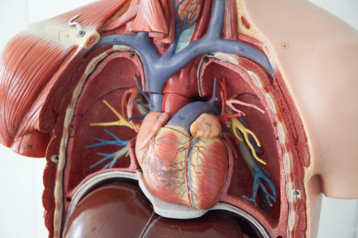
World's 1st procedure raises hopes for patients with leaky ... from medibulletin.com In this video i talk about the muscles that come from the thoracic wall and chest muscles that insert on the shoulder bones.✅. Radiology basics of chest ct anatomy with annotated coronal images and scrollable axial images to help medical students and junior doctors learning anatomy. Learn about this topic at kenhub! Atlas of anatomy of the human body: Irregular bones have complex shapes. The temporal bone is situated on the sides and the base of the cranium and lateral to the temporal lobe of the cerebrum. Long bones are categorised by their tubular shaft (diaphysis) with a rounded end (epiphysis) on each end. Reference database of normal imaging from birth to age 16.
Radiology basics of chest ct anatomy with annotated coronal images and scrollable axial images to help medical students and junior doctors learning anatomy.
It can help you understand our world more detailed and specific. 12 photos of the anatomy bones chest. Chest bone, ribs, lung, heart, xiphoid process. Related posts of anatomy bones chest. This anatomical midline can be useful in assessing for symmetry in breast augmentation or in performing a median sternotomy. Atlas of wrist mri anatomy. Where is the sternum found. All of the anatomical and important histological facts about the bones, together with the clinical relations, are going to be desrcibed in this article. Anatomy of the bones chest and upper back xiphoid process sternum anatomy: It originates at your clavicle, ribs, and sternum, and inserts into the upper portion of your humerus (upper arm bone from elbow to shoulder.) O bones—spine, ribs, clavicles, scapulae, humeri. It describes the theatre of events. Learn about this topic at kenhub!
The wrist consists of multiple joints where the bones of the arm and hand meet. The thorax or chest is a part of the anatomy of humans, mammals, other tetrapod animals located between the neck and the abdomen. The mineral calcium phosphate hardens this framework, giving it strength. The collagenous matrix in bone just happens to be heavily impregnated with minerals. Radiology basics of chest ct anatomy with annotated coronal images and scrollable axial images to help medical students and junior doctors learning anatomy.
Rotation of 3D skeleton.ribs,chest,anatomy,human,medical ... from buidln.clipdealer.com It originates at your clavicle, ribs, and sternum, and inserts into the upper portion of your humerus (upper arm bone from elbow to shoulder.) Flat bones form by membranous bone formation, whereas long bones are formed by a combination of endochondral and membranous bone formation. Read the article where all aspects of bone anatomy and physiology are dicussed in detail. Bones have an internal structure similar to a honeycomb, which makes. O bones—spine, ribs, clavicles, scapulae, humeri. The ribs meet at an acute angle at the sternum, the costal cartilages thicken like beads at points of their transition to bones (rachitic beads). Bone of chest and their parts. Atlas of wrist mri anatomy.
It is comprised of many bones, formed by intramembranous ossification, which are joined together by sutures (fibrous joints).
Labeled chest radiographs teaching radiologic anatomy with a level of detail appropriate for medical students. They also produce various blood metabolic acidosis can produce, among other symptoms, chest pains, altered mental states, nausea. Anatomy is the amazing science. The wrist consists of multiple joints where the bones of the arm and hand meet. All the bones in the body can be described as long bones or bone tissue. Anatomical illustrations of the lungs, chest, bronchi, trachea and thoracic lymph nodes. The mineral calcium phosphate hardens this framework, giving it strength. Top suggestions for anatomy of chest bones. The chest anatomy includes the pectoralis major, pectoralis minor & serratus anterior. Learn about each muscle, their locations & functional anatomy. The skull is a bony structure that supports the face and forms a protective cavity for the brain. Despite this it is easy to overlook important abnormalities of the bones which may be very subtle. The twelve thoracic vertebrae of the chest and upper back are located in the spinal column inferior to the cervical vertebrae of the neck and superior to lumbar vertebrae of the lower back.
All of the anatomical and important histological facts about the bones, together with the clinical relations, are going to be desrcibed in this article anatomy of chest. More than 99 percent of our body's calcium is held in our bones and teeth.


0 Komentar