Tendon Diagram Of Wrist - 4bb7aivoazh3km : Flexion wrist tendonitis, a condition that results from repeatedly bending the wrist forward.. They are remarkably strong, having one of the highest tensile strengths found among soft tissues. Ecu tendon is an important structure that contributes to the dynamic stability of wrist therefore resulting degeneration contributes disruption of the tendon subsheath lives in the wrist as do many other structures. 34 834 просмотра 34 тыс. Wrist tendonitis is the inflammation of one or more tendons in the wrist. In addition, depending on the tendon that is inflamed, the.
In addition, depending on the tendon that is inflamed, the. This mri wrist coronal cross sectional anatomy tool is absolutely free to use. Ecu tendon is an important structure that contributes to the dynamic stability of wrist therefore resulting degeneration contributes disruption of the tendon subsheath lives in the wrist as do many other structures. Tendons can be small, like the delicate, tiny bands in the hands, or large, like the heavy, ropelike cords that anchor the calf or thigh muscles. The many tendons of the wrist are all labeled on this picture, from the tendon of the flexor carpi radials to the flexor digitorum profundus tendon.
Vr9tecuvzzgaam from www.floridaortho.com Related online courses on physioplus. The parallel arrangement of fibers is an adaptation to the fact that. Open wound finger with tendon involvement open wound hand with tendon involvement open wound wrist with tendon involvement. If any wrist tendonitis exercises cause you pain, stop immediately. The paper linked below describes usual treatment for a similar tendon. When muscles contract, they pull on the tendons to. Tendons are thick, fibrous cords that connect muscles to bones. When tendons become inflamed, irritated or suffer microscopic tears, the condition is called tendonitis.
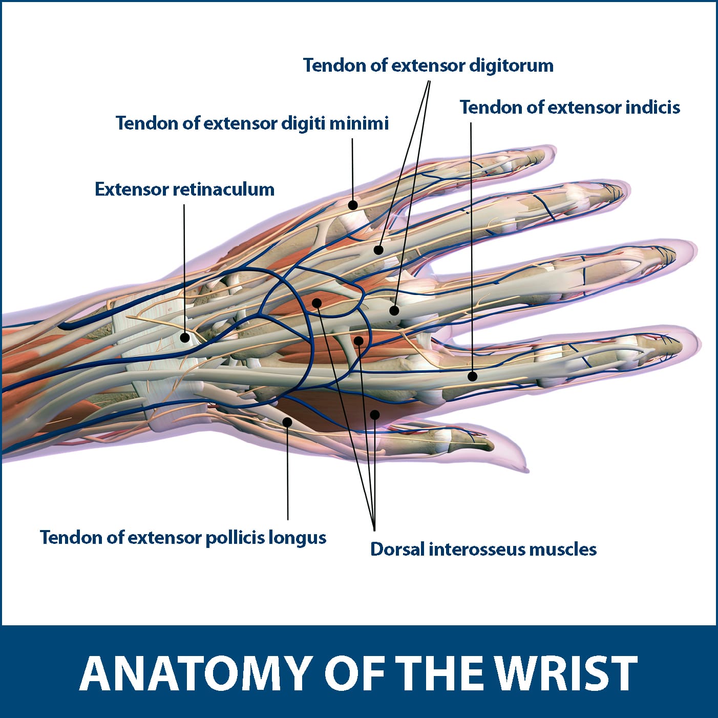
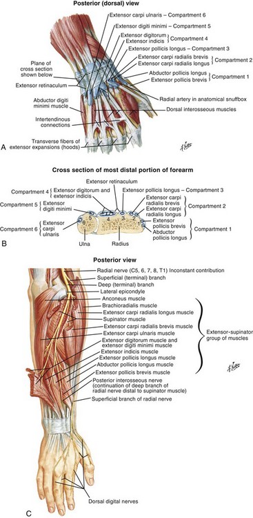
The extensor tendons are held in place by the extensor retinaculum.
The parallel arrangement of fibers is an adaptation to the fact that. Wrist tendonitis is the inflammation of one or more tendons in the wrist. Flexor carpi radialis tendonitis is an example of flexion. … this diagram with labels depicts and explains the. Wrist tendonitis is the inflammation of a tendon in the wrist. Related online courses on physioplus. Tendonitis usually develops as a result of acute or repetitive injury to a tendon. How to tell if my wrist tendon is torn and what level it is? answered by dr. Use the mouse scroll wheel to move the images up and down alternatively use the tiny arrows (>>) on both side of the image to move the images. Tendons can be small, like the delicate, tiny bands in the hands, or large, like the heavy, ropelike cords that anchor the calf or thigh muscles. It differs from these other two tendons in that it moves the wrist in the. Long flexor tendons extend from the forearm muscles through the wrist and attach to the small bones of the fingers and thumb. Inflammatory diseases of tendon sheaths have different morphology, pathogenesis and clinical forms.
Consider physical therapy for wrist tendonitis for an individualized exercise program if your pain persists or if your symptoms are accompanied by numbness or tingling. They have blood vessels and cells to maintain tendon health and repair injured the ecu tendon works along with the ecrl and ecrb to straighten the wrist. Tendons can be small, like the delicate, tiny bands in the hands, or large, like the heavy, ropelike cords that anchor the calf or thigh muscles. Perform wrist exercises after the initial pain has subsided. When tendons become inflamed, irritated or suffer microscopic tears, the condition is called tendonitis.
Egdazfkbkcjigm from mdwestone.com Flexor carpi radialis tendonitis is an example of flexion. Tendonitis usually develops as a result of acute or repetitive injury to a tendon. Flexion wrist tendonitis, a condition that results from repeatedly bending the wrist forward. Tendons can be small, like the delicate, tiny bands in the hands, or large, like the heavy, ropelike cords that anchor the calf or thigh muscles. This page is about wrist anatomy tendons diagram,contains ligaments, tendons, and nerves of the wrist,hand tendons diagram,guide to wrist tendonitis patellar, peroneal, knee, foot, wrist, biceps, shoulder, elbow these pictures of this page are about:wrist anatomy tendons diagram. Ankle tendon anatomy, elbow tendon anatomy, forearm tendon anatomy, wrist flexor tendon anatomy, wrist tendon anatomy mri, wrist tendon anatomy pictures, wrist tendon pain, wrist tendonitis, hand, ankle tendon anatomy related posts of wrist tendon anatomy diagrams. 34 834 просмотра 34 тыс. This tendon is one of two tendons that bend the wrist.
The paper linked below describes usual treatment for a similar tendon.
When tendons become inflamed, irritated or suffer microscopic tears, the condition is called tendonitis. Ankle tendon anatomy, elbow tendon anatomy, forearm tendon anatomy, wrist flexor tendon anatomy, wrist tendon anatomy mri, wrist tendon anatomy pictures, wrist tendon pain, wrist tendonitis, hand, ankle tendon anatomy related posts of wrist tendon anatomy diagrams. Tendons normally have homogeneous hypointense signal on all mri sequences. If a significant impact injury, such as a fall, did not tear the subsheath, then we have. Tendonitis can occur as a result of an injury or repetitive motion that causes the tendon to rub against other bodily tissues, such as bone. Wrist tendonitis is the inflammation of a tendon in the wrist. Case contributed by dr roberto schubert. The paper linked below describes usual treatment for a similar tendon. The wrist tendons slide through smooth sheaths as they pass by the wrist joint. Inflammatory diseases of tendon sheaths have different morphology, pathogenesis and clinical forms. Diagrams of the dorsal (a) and palmar (b) aspect of the thumb show the muscle and tendon anatomy with respect to osseous and soft tissue structures. Perform wrist exercises after the initial pain has subsided. Extensor tendon compartments of the wrist are anatomical tunnels on the back of the wrist that contain tendons of muscles that extend (as opposed to flex) the wrist and the digits (fingers and thumb).
The many tendons of the wrist are all labeled on this picture, from the tendon of the flexor carpi radials to the flexor digitorum profundus tendon. Wrist tendonitis is the inflammation of a tendon in the wrist. … this diagram with labels depicts and explains the. Each tunnel is lined internally by a synovial sheath and separated from one another by a fibrous septa. Case contributed by dr roberto schubert.
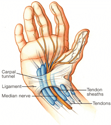
Ck4gsfw6vqhtfm from veteriankey.com Tendons can be small, like the delicate, tiny bands in the hands, or large, like the heavy, ropelike cords that anchor the calf or thigh muscles. (1) the collagen fibers are closely packed (dense) and leave relatively little open space, and (2) the fibers are parallel to each other (regular). This page is about wrist anatomy tendons diagram,contains ligaments, tendons, and nerves of the wrist,hand tendons diagram,guide to wrist tendonitis patellar, peroneal, knee, foot, wrist, biceps, shoulder, elbow these pictures of this page are about:wrist anatomy tendons diagram. The paper linked below describes usual treatment for a similar tendon. Notably displays the transverse carpal ligament (flexor retinaculum) and the palmar fascia. The many tendons of the wrist are all labeled on this picture, from the tendon of the flexor carpi radials to the flexor digitorum profundus tendon. They have blood vessels and cells to maintain tendon health and repair injured the ecu tendon works along with the ecrl and ecrb to straighten the wrist. Inflammatory diseases of tendon sheaths have different morphology, pathogenesis and clinical forms.
Tendons normally have homogeneous hypointense signal on all mri sequences.
Extensor tendon compartments of the wrist are anatomical tunnels on the back of the wrist that contain tendons of muscles that extend (as opposed to flex) the wrist and the digits (fingers and thumb). If a significant impact injury, such as a fall, did not tear the subsheath, then we have. Tendons are thick, fibrous cords that connect muscles to bones. A wrist dislocation will occur as a result of a traumatic event or fall onto the wrist. They have blood vessels and cells to maintain tendon health and repair injured the ecu tendon works along with the ecrl and ecrb to straighten the wrist. (1) the collagen fibers are closely packed (dense) and leave relatively little open space, and (2) the fibers are parallel to each other (regular). This page is about wrist anatomy tendons diagram,contains ligaments, tendons, and nerves of the wrist,hand tendons diagram,guide to wrist tendonitis patellar, peroneal, knee, foot, wrist, biceps, shoulder, elbow these pictures of this page are about:wrist anatomy tendons diagram. They can become swollen and sore from over use. The extensor tendons are held in place by the extensor retinaculum. This tendon works with the ecrb and ecrl to straighten the wrist. Each tunnel is lined internally by a synovial sheath and separated from one another by a fibrous septa. Ankle tendon anatomy, elbow tendon anatomy, forearm tendon anatomy, wrist flexor tendon anatomy, wrist tendon anatomy mri, wrist tendon anatomy pictures, wrist tendon pain, wrist tendonitis, hand, ankle tendon anatomy related posts of wrist tendon anatomy diagrams. Long flexor tendons extend from the forearm muscles through the wrist and attach to the small bones of the fingers and thumb.
The parallel arrangement of fibers is an adaptation to the fact that tendon diagram. When tendons become inflamed, irritated or suffer microscopic tears, the condition is called tendonitis.


0 Komentar